The Human Eye: A Complex and Fascinating Organ
The human eye is a remarkable and intricate organ that enables us to perceive the world around us. It’s essential to take care of our eyes, protecting them from dust, foreign objects, and injury. In this article, we’ll delve into the various parts of the eye and their functions.
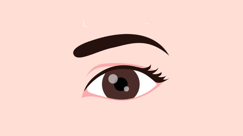
Eyelids: The Guardians of the Eye
Eyelids are the first line of defense for our eyes. These flaps open and shut voluntarily, but also have involuntary actions like blinking to retain moisture and closing reflexively to protect the eye from harm. The orbicularis oculi muscle surrounds the eye, controlling eyelid movement.
Tear Gland: Lubricating the Eye
The lacrimal gland produces tears, which spread across the eye’s surface with each blink. Tears lubricate, moisturize, and wash away infection-causing germs and dust particles. Excess tears drain through the lacrimal ducts into the nose.
Sclera: The Protective Outer Layer
The sclera is the white, tough outer wall of the eye, protecting the internal parts. It’s covered by a thin, transparent conjunctiva tissue. Blood vessels visible in the white part of the eye belong to the sclera and conjunctiva.
Iris: The Colorful Controller
The iris is the colored ring around the pupil, controlling the amount of light entering the eye. Muscles in the iris expand and contract to adjust the pupil’s size, focusing light on the object being observed.
Pupil: The Light Regulator
The pupil is the dark spot at the center of the iris, changing size to regulate light entry. The iris muscles control the pupil’s size, ensuring optimal light levels for clear vision.
Cornea: The Clear Surface
The cornea is the clear, convex surface at the front of the eye, helping focus light rays before they enter the eye. It’s a crucial part of the eye’s refractive system.
Lens: The Focusing Element
The lens is an elliptical structure behind the iris, sharpening focus on the object being observed. Its curvature focuses light on the retina, enabling clear vision.
Vitreous Cavity: The Gel-Like Filling
The vitreous cavity is the space between the lens and the eyeball’s back, filled with a clear, jelly-like substance called vitreous. This fluid helps maintain the eyeball’s shape.
Retina: The Light-Sensitive Layer
The retina is a thin layer of tissue lining the eyeball’s inner wall, containing millions of nerve endings that capture visual images. When light hits the retina, these nerve endings convert it into electrical impulses, transmitted to the brain via the optic nerve.
Photoreceptor Cells: Converting Light to Signals
The retina contains two types of photoreceptor cells: rods and cones. These cells convert light into electrochemical signals, enabling us to perceive the world around us.
Retinal Pigment Epithelium (RPE): Supporting the Photoreceptors
The RPE is a layer of tissue beneath the photoreceptors, absorbing excess light for clearer vision. It also transports nutrients and waste in and out of the eye.
Choroid: Supplying Blood and Nutrients
The choroid is the structure behind the retina, supplying blood and nutrients to the retina and RPE.
Macula: The Detail-Focused Area
The macula is a dense collection of light-sensitive cells at the retina’s center, enabling us to see fine details.
Optic Nerve: Transmitting Visual Signals
The optic nerve carries electrical impulses from the retina to the brain, where they’re interpreted and identified.
Muscles of the Eyeball: Controlling Movement
Six muscles attached to the sclera control the eyeball’s movement, enabling us to track objects without excessive head movement.
References:
https://www.mayoclinic.org/healthy-lifestyle/adult-health/multimedia/eyes/sls-20076536?s=7